On 18th February, there was an award ceremony for the Virtual Research and Innovation Exhibition (EREKA 2021) organized by Universiti Malaysia Perlis (UniMAP) which was conducted virtually.
EREKA 2021 has been conducted virtually on three phases, Registration on 18th to 30th January, Online Judging on 31st January to 3rd February, and Award Ceremony on 18th February.
INEE sent eight entries from research studies from INEE researchers to EREKA 2021, where all entries have been awarded medals and one of them received Best of The Best Award.
Here are eight products with title and researchers who received the EREKA 2021 award:
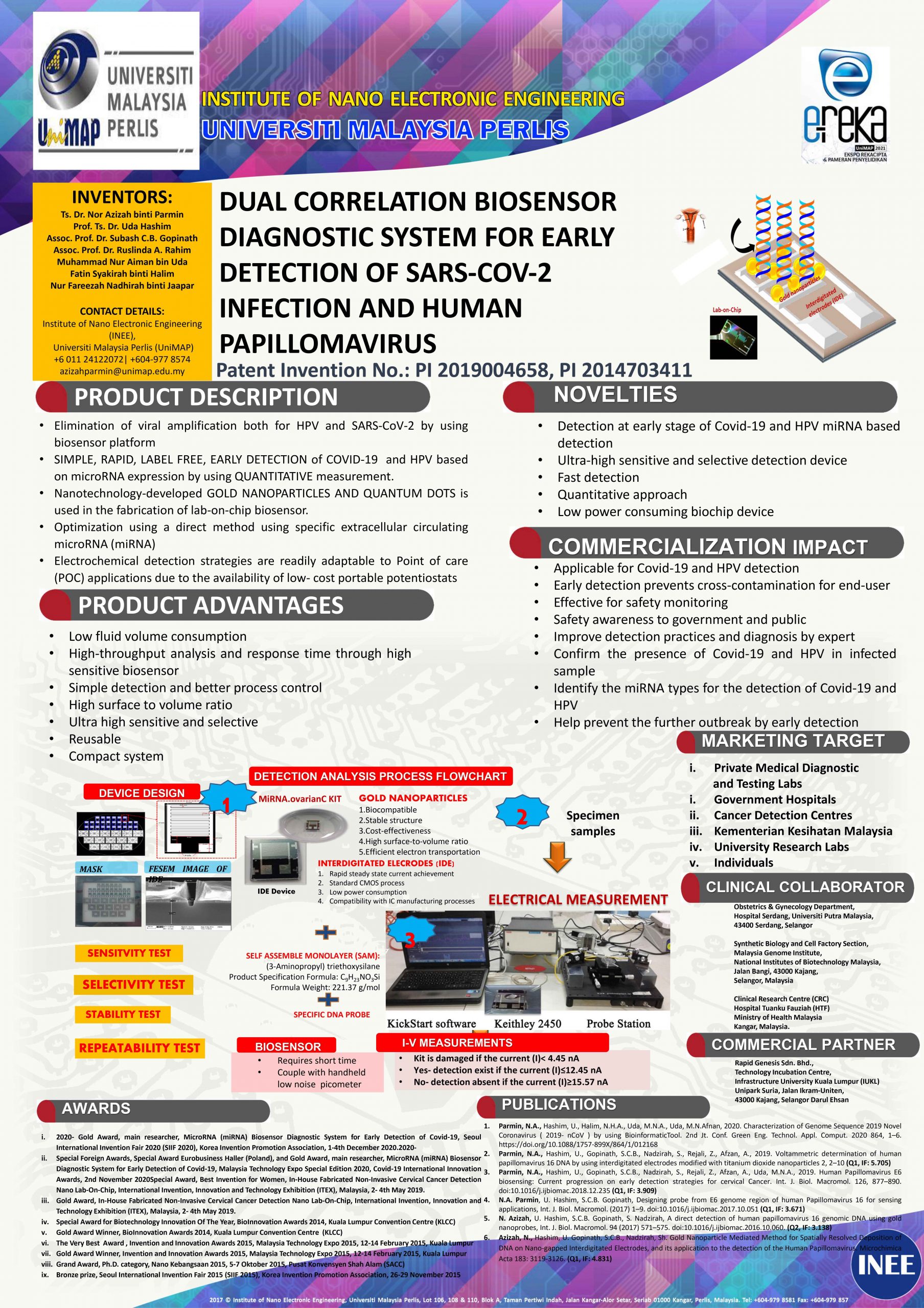
Best of The Best Award
Researcher: Ts. Dr. Nor Azizah Parmin
Title: Dual Correlation Biosensor Diagnostic System for Early Detection of COVID-19 Infection and Human Papillomavirus
Gold Medal
Researcher: Prof. Ts. Dr. Uda Hashim
Title: 12C19 E-Detector
Researcher: Assoc. Prof. Dr. Mohammad Nuzaihan Md Nor
Title: Modified Silicon Nanostructures as Diabetes Mellitus Sensor
Researcher: Dr. Mohamad Faris Mohamad Fathil
Title: Field-Effect Device for AMI Biomarker Detection
Researcher: Ts. Dr. Nor Azizah Parmin
Title: Dual Correlation Biosensor Diagnostic System for Early Detection of COVID-19 Infection and Human Papillomavirus
Researcher: Ts. Dr. Nor Azizah Parmin
Title: A Sensitive MicroRNA (MiRNA) Biosensor Based on Carbon Quantum Dots (CQD) for Early Detection of COVID-19
Researcher: Ts. Dr. Nor Azizah Parmin
Title: Multi Detection of Genosensor for Human Papillomavirus (HPV) Strains 16, 18, and 58 for Early Detection of Cervical Cancer
Silver Medal
Researcher: Assoc. Prof. Dr. Subash C B Gopinath
Title: Archimedean NanoMIP Sensor: A Prognostic Device for Clotting Disease Detection
Researcher: Assoc. Prof. Dr. Ruslinda Abdul Rahim
Title: Graphene-modified Electrode for Detection of BPA
Congratulations to all the research teams that participated in the EREKA 2021.
Best of The Best Award Poster